


產(chǎn)品詳情
擬人化
被鑄造的Acround頭骨
檢查重要的物理參數(shù)
提供真實(shí)的系統(tǒng)檢查
理想的培訓(xùn)
聚碳酸酯底座的尺寸適用于與縱軸平行或垂直的掃描
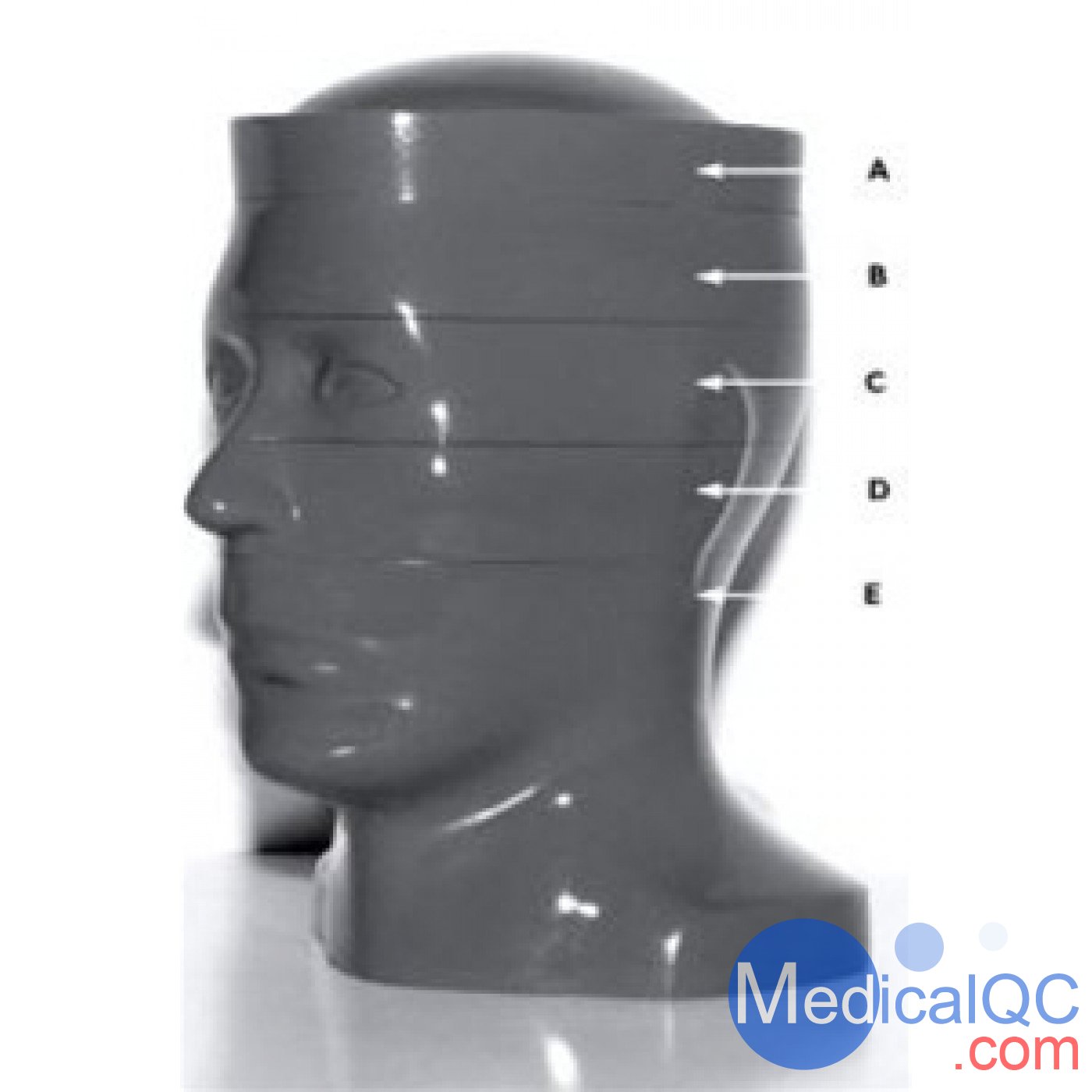
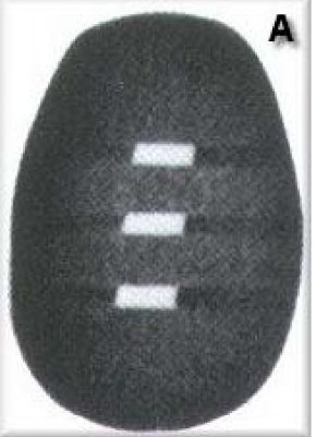
A部分: 0.4毫米厚的鋁板設(shè)置45°角,測(cè)量光束寬度和切片厚度。
RS-250 A部分
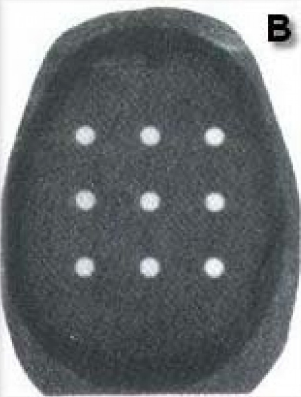
B部分:劑量測(cè)定部分提供患者暴露控制,并在實(shí)際患者掃描前檢查異常內(nèi)部劑量。定制容納TLD與棒或芯片是可用的。
RS-250 B部分
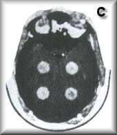
C部分:分級(jí)大小和放射性的圓柱形腫瘤建立高對(duì)比度和低對(duì)比度分辨率,并顯示部分光束平均效應(yīng)。
RS-250 C部分
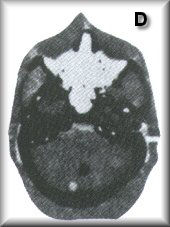
D部分:擬人部分在空氣對(duì)比的內(nèi)耳和骨折的巖骨中提供實(shí)際的聽(tīng)覺(jué)小骨。后顱窩小腫瘤被放置在大多數(shù)掃描儀難以成像的區(qū)域。
RS-250 D部分
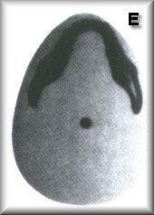
E部分:人體模型縱軸上1/4英寸直徑的鋁棒,檢查一般對(duì)準(zhǔn)情況,“干水”給出了一個(gè)現(xiàn)實(shí)的“噪音檢查”。
CT Head with 5 slices to test all beters of computerized tomography
Anthropomorphic
Molded Around Skull
Checks Important Physical Parameters
Provides Realistic System Check
Ideal For Training
Polycarbonate Base Dimensioned For Scans Parallel or Perpendicular to Longitudinal Axis
SECTION A: Aluminum plates, 0.4mm thick, set at a 45° angle, measure beam b and slice thickness.
SECTION B: The dosimetry section provides patient-exposure controls, and checks on abnormal internal doses before actual patient scans. Customization to accommodate TLD with rods or chips is available.
SECTION C: Cylindrical tumors of graded sizes and radiodensities establish high and low-contrast resolution and demonstrate partial beam-averaging effects.
SECTION D: The anthropomorphic section provides actual auditory ossicles in an air-contrast inner ear and a fractured petrous bone. Small tumors in the posterior fossa are placed in an area which most scanners image with difficulty.
SECTION E: 1/4 Inch diameter aluminum rod on the phantom's longitudinal axis, checks general alignment, and "DryWater" gives a realistic "noise check."
SAG:RS-250,RSD RS-250,RS-250頭部CT模體,頭部CT模體,RS-250 CT模體
